Dye-loaded polymeric nanoparticles
In 2014, we proposed a new concept of fluorescent polymer NPs, doped with ionic liquid-like salts of a cationic dye (octadecyl rhodamine B) with a bulky hydrophobic counterion (fluorinated tetraphenylborate).1 The latter serves as spacer between cationic dyes minimizing dye aggregation and self-quenching (Figure 1). Remarkably, obtained ultra-bright nanoparticles undergo due to efficient dye-dye communication, the particles undergo photo-induced reversible on/off fluorescence switching (blinking).1 This implies collective on off behavior of >100 dyes coupled by fast excitation energy transfer. The size of particles can be tuned down to 15 nm using single negative charge in polymer (PMMA, PLGA or PCL).2 The obtained 15-nm PMMA-based NPs loaded with rhodamine based dyes are >10-fold brighter than quantum dots (Figure 1).2 Moreover, by tuning dye loading and polymer matrix we can generate nanoparticles presenting stable emission or complete on/off switching(blinking).3 Using photochromic dyes, these NPs can be effectively photo-switched.4
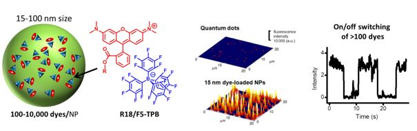
Figure 1. Design of dye-doped fluorescent NPs using bulky counterions (left); fluorescence microscopy images of quantum dots QD585 and 15 nm PMMA-MA NPs containing R18/F5-TPB immobilized on a glass surface (middle): our NPs are >10-fold brighter than QDs; on/off switching (blinking) of single PLGA NPs containing >100 dyes (R18/F5-TPB) per particle. Data adapted from.1,2
These NPs, being spontaneously endocytosed by living cells, feature high signal-to-noise ratio and absence of toxicity. The counterion-based concept opens the way to a new class of ultrabright nanomaterials for sensing, imaging and light harvesting. This class of nanoparticles has been recently reviewed by us.5
Giant light-harvesting nanoantenna for single molecule detection
Nanoparticles encapsulating large number of dyes could serve as light harvesting antenna that collect light energy by ensemble of donor dyes and transfer to single dye acceptors, as realized by nature in photosynthetic centers (chloroplasts). Recently, we showed than our 60 nm PMMA polymer nanoparticles containing >10,000 rhodamine-based donor dyes can efficiently transfer energy to 1–2 acceptors.6 As a result the emission of Cy5-based acceptor dye is amplified ~1,000 (Figure 2) and it becomes 25-fold brighter than quantum dots QD655. This unprecedented amplification of the acceptor dye emission enables observation of single molecules at illumination powers (1–10 mW cm-2) that are >10,000-fold lower than typically required in single-molecule measurements. Finally, using a basic set-up, which includes a ×20 air objective and a sCMOS camera, we could detect single Cy5 molecules by simply shining divergent light on the sample at powers equivalent to sunlight.6
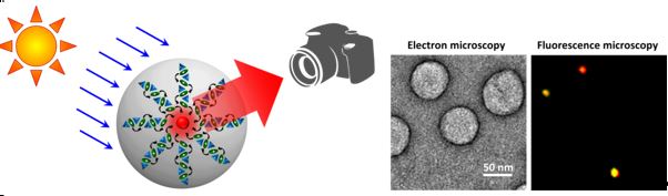
Figure 2. Scheme of nanoparticle antenna containing >10,000 donor dyes and 1-2 acceptor dyes, which can be detected by simple imaging setup using illumination equivalent to sunlight. Right: electron microscopy image of 60-nm nanoantennas and fluorescence photographs of single acceptor dye molecules inside nanoantenna using sunlight mimicking conditions. Data adapted from.6
Cell barcoding
An important application of fluorescence nanoparticles is long-term cell tracking. Recently we developed nanoparticles of three different color built from biodegradable poly(lactic-co-glycolic acid) polymer loaded with cyanine dyes (DiO, DiI, and DiD) with the help of bulky fluorinated counterions.5 Mixing nanoparticles of three colors in different proportions generates a homogeneous RGB (red, green, and blue) barcode in cells, which is transmitted through many cell generations (Figure 3). Cell barcoding has already been validated on 7 cell lines, 13 color codes, and it enables simultaneous tracking of co-cultured barcoded cell populations for >2 weeks.7 This technology enabled six-color imaging in vivo for tracking xenografted cancer cells and monitoring morphogenesis after microinjection in zebrafish embryos (Figure 3).7 In addition to a robust method of multicolor cell labeling and tracking, this work suggests that multiple functions can be co-localized inside cells by combining structurally close nanoparticles carrying different functions.
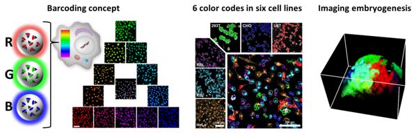
Figure 3. Cell barcoding in vitro and in vivo using combination of cyanine-loaded polymer NPs of different color. The large micrograph shows a confocal image of six mixed cell types labeled with different RGB barcodes. Imaging of the development of zebrafish embryos labeled by intracellular injection of fluorescent NPs. Data adapted from.7
References
1) Reisch, A., P. Didier, L. Richert, S. Oncul, Y. Arntz, Y. Mely and A.S. Klymchenko, Collective fluorescence switching of counterion-assembled dyes in polymer nanoparticles. Nature Communications, 2014, 5, 4089.
2)Reisch, A., A. Runser, Y. Arntz, Y. Mely and A.S. Klymchenko, Charge-Controlled Nanoprecipitation as a Modular Approach to Ultrasmall Polymer Nanocarriers: Making Bright and Stable Nanoparticles. ACS Nano, 2015, 9, 5104-5116.
3) Reisch, A., Trofymchuk, K., Runser, A., Fleith, G., Rawiso, M., and Klymchenko, A.S. Tailoring Fluorescence Brightness and Switching of Nanoparticles through Dye Organization in the Polymer Matrix ACS Appl. Mater. Interfaces, DOI: 10.1021/acsami.7b12292.
4) Trofymchuk K., Prodi L., Reisch A., Mély Y., Altenhöner K., Mattay J., Klymchenko A.S. Exploiting fast exciton diffusion in dye-doped polymer nanoparticles to engineer efficient photoswitching, J. Phys. Chem. Lett. 2015, 6, 2259−2264.
5) Reisch, A. and A.S. Klymchenko, Fluorescent Polymer Nanoparticles Based on Dyes: Seeking Brighter Tools for Bioimaging. Small, 2016, 12, 1968-1992.
6) Trofymchuk, K.; Reisch, A.; Didier, P.; Fras, F.; Gilliot, P.; Mely, Y.; Klymchenko, A. S.: Giant light-harvesting nanoantenna for single-molecule detection in ambient light. Nature Photonics 2017, 11, 657.
7) Andreiuk, B., A. Reisch, M. Lindecker, G. Follain, N. Peyrieras, J.G. Goetz and A.S. Klymchenko, Fluorescent Polymer Nanoparticles for Cell Barcoding In Vitro and In Vivo. Small, 2017, 13(38).
Dye-loaded lipid nanoparticles
Lipid nanoparticles are particularly interesting for bioimaging applications because they are composed of fully biodegradable materials and are very easy to fabricate by spontaneous emulsification. To ensure efficient dye loading with minimal leakage from the particles, we followed successfully two concepts of dye design: (i) dyes bearing multiple hydrophobic alkyl chains1 and (ii) cationic cyanine dyes in combination with bulky hydrophobic counterions.2
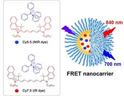 |
Figure 1. Chemical structures of lipophilic cyanine 5.5 and 7.5 dyes (Cy5.5LP and Cy7.5LP) with their bulky hydrophobic counterions and schematic presentation of nano-droplet encapsulating these dyes. |
By using bulky hydrophobic counterion tetraphenyl borate (TPB), we were able to increase many-fold the solubility of cyanines dyes in oil and create 90 nm lipid droplets bearing exceptional number of dyes (~ 12,000 per droplet, 8 wt% dye loading). The extreme brightness of these NCs (100-fold brighter than quantum dots) enabled to pioneer single particle tracking in vivo in a zebrafish embryo model. Further, using this concept, we have developed FRET nanocarriers loaded with NIR dyes Cy5.5 Cy7.5 (Figure 1).3 Efficient FRET and excellent brightness of the loaded dyes allowed achieving whole-animal imaging with a very high resolution (Figure 2). Moreover, we were able to accurately resolve the follow-up of nanoparticles integrity directly in the blood circulation of living mice (Figures 2, 3).3 It was found that lipid nanocarriers despite their liquid core can enter tumors in nearly intact form, showing their capacity to deliver and release their inside target tumors.
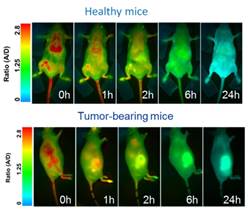
Figure 2. FRET imaging of healthy and nude mice at different times after injection with NIR-FRET lipid NCs (1% of Cy5.5LP and Cy7.5LP each). |
Figure 3. Concept of FRET NCs that can report on their integrity by change in their emission color. |
References
1) Klymchenko, A.S.; Roger, E.; Anton, N.; Anton, H.; Shulov, I.; Vermot, J.; Mely, Y.; Vandamme, T.F. Highly lipophilic fluorescent dyes in nano-emulsions: towards bright non-leaking nano-droplets. RSC Advances, 2012, 2, 11876-11886.
2) Kilin, V.N., H. Anton, N. Anton, E. Steed, J. Vermot, T.E. Vandamme, Y. Mely and A.S. Klymchenko, Counterion-enhanced cyanine dye loading into lipid nano-droplets for single-particle tracking in zebrafish. Biomaterials, 2014. 35(18): p. 4950-4957.
3) Bouchaala, R., N. Anton, H. Anton, T. Vandamme, J. Vermot, D. Smail, Y. Mely and A.S. Klymchenko, Light-triggered release from dye-loaded fluorescent lipid nanocarriers in vitro and in vivo. Colloids and Surfaces B-Biointerfaces, 2017. 156: p. 414-421.
Protein-sized nanoparticles
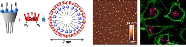
Figure 1. PEGylated cyanine bis-azides form a covalently attached “corona” on micelles of amphiphilic calixarene bearing alkyne groups. AFM image of CX/Cy3L micelles in water and fluorescence confocal image of HeLa cells incubated with micelles for 3 h. Green corresponds to plasma membrane staining with WGA-Alexa Fluor488 (50 nm), while red (donor channel) and magenta (FRET channel) correspond to the micelles.
The key challenge in the field of fluorescent nanoparticles (NPs) for biological applications is to achieve superior brightness for sizes equivalent to single proteins (3–7 nm). Recently, we have developed protein-sized (7-nm) ultrabright fluorescent nanoparticles, in which PEGylated cyanine 3 and 5 bis-azides form a covalently attached corona on micelles of amphiphilic calixarene bearing four alkyne groups (Figure 1). In the ON-state, the obtained NPs are ~2-fold brighter than quantum dots (QD-585), which makes them the smallest PEGylated organic NPs of this high brightness. FRET studies suggest that they are highly stable in aqueous and organic media as well as in living cells, in which they show excellent imaging contrast.
Another type of our developed small ultrabright fluorescent micelles relay on self-assembling of specially designed cationic dye amphiphiles. We showed that a non-coordinating anion, fluorinated tetraphenylborate, assembles cationic cyanine amphiphiles into 7–8 nm fluorescent nanoparticles that are 40-fold brighter than a single cyanine dye (Figure 2). They exhibited efficient FRET to a single acceptor as well as high stability.
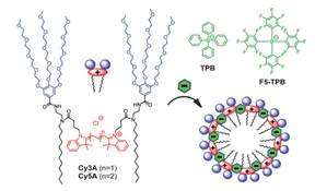
Figure 2. Cyanine amphiphiles and their assembly into micelles in the presence of non-coordinating anions.
References
1) Shulov, I., Y. Arntz, Y. Mely, V.G. Pivovarenko and A.S. Klymchenko, Non-coordinating anions assemble cyanine amphiphiles into ultra-small fluorescent nanoparticles. Chemical Communications, 2016. 52(51): p. 7962-7965.
2) Shulov, I., R.V. Rodik, Y. Arntz, A. Reisch, V.I. Kalchenko and A.S. Klymchenko, Protein-Sized Bright Fluorogenic Nanoparticles Based on Cross-Linked Calixarene Micelles with Cyanine Corona. Angewandte Chemie-International Edition, 2016. 55(51): p. 15884-15888.